Optical genome mapping (OGM) is a method for the identification of structural variants (SVs) in the genome that closes gaps in existing diagnostics. It enables high-resolution detection of SVs, including balanced translocations and inversions, with a resolution of up to 1 kb. OGM is particularly useful for breakpoint characterization of balanced SVs, analysis of complex chromosomal rearrangements and detection of submicroscopic SVs in patients with developmental delay, intellectual disability or malformations after negative conventional diagnostics.
Structural variants–defined as differences of at least 50 base pairs (bp) between an individual genome and the human genome reference–represent a significant source of human diversity. While single nucleotide variants (SNVs) have higher prevalence in the genome, SVs can exert a stronger impact on the genome and phenotypes due to the alteration of many nucleotides in a single event. Additionally, SVs are more likely to affect coding regions and involve more than one gene. Next-generation sequencing (NGS) technology is the method of choice for detecting SNVs and smaller, intragenic copy number variations (CNVs) such as deletions or duplications. However, the detection of larger CNVs or balanced SVs such as translocations or inversions remains a significant challenge.
The methods currently used in cytogenetic routine diagnostics each have specific advantages and limitations. Conventional chromosome analysis allows the detection of both unbalanced and balanced SVs at the single-cell level, but only offers a low resolution of 5–10 megabase pairs (Mb). In contrast, chromosomal microarray analysis (CMA) enables high-resolution detection of CNVs as small as approximately 10 kilobase pairs (kb) but cannot detect balanced SVs.
An elegant approach to bridge this detection gap is optical genome mapping (OGM, next-generation cytogenomics, next-generation mapping).
This method, first developed in the early 2010s, involves isolating ultra-high molecular weight DNA in fragments ranging from 100 kb to 2 Mb. A fluorescence labeling process creates a specific banding pattern by targeting a recognition sequence that occurs over 500,000 times in the haploid genome. After linearization of the DNA fragments in nanofluid channels, the banding pattern is scanned using a laser (compare this with conventional chromosome analysis, which typically achieves an average of 500–600 bands). Bioinformatic comparison between the patient and reference genome banding patterns then enables the detection of SVs with a resolution of up to 1 kb.
Optical genome mapping is an important addition to existing diagnostics for patients with developmental delays, intellectual disabilities, or congenital anomalies. Current stepwise diagnostics for this patient cohort – comprising conventional chromosome analysis (detection rate up to 15%), CMA (approximately 20%), and panel/exome analysis (20–25%) – leave around 40% of cases undiagnosed. In 2020, Shieh et al. demonstrated the potential of optical genome mapping by identifying pathogenic SVs in 4 out of 23 patients with negative exome analysis. Similarly, Mantere et al. showed in their 2020 study involving 85 patients with 100 known structural and numerical chromosomal aberrations (detected via chromosome analysis or CMA) the high diagnostic value of optical genome mapping. Not only were all 100 aberrations identified, but the breakpoints of the structural SVs were also fully characterized. An internal validation study of 16 patients further confirmed that all aberrations were detected via optical genome mapping, with breakpoints of balanced SVs pinpointed to regions ranging from 11 kb to 800 bp.
Indications for the Application of Optical Genome Mapping:
- Characterization of breakpoints in balanced SVs (e.g., translocations, inversions).
- Analysis of marker chromosomes and complex chromosomal rearrangements.
- Characterization of duplications (e.g., tandem vs. insertion, inverted vs. non-inverted).
- Detection of submicroscopic SVs in patients with developmental delays, intellectual disabilities, or anomalies after negative stepwise diagnostics.
- Identification of SVs in the second allele in autosomal recessive disorders after detecting an SNV in the first allele.
References
Ho et al. 2020, Nat Rev Genet 21:171 / Li et al. 2017, Genome Bio 18:230 / Lindstrand et al. 2019, Genome Med 11:68 / Mantere et al. 2020, bioRxiv doi.org/10.1101/ 2020.07.15.205245 / Mostovoy et al. 2020, bioRxiv, doi.org/10.1101/2020.04.30.071449 / Shieh et al. 2021, NPJ Genom Med 6:77 / Wang et al. 2020, J Assist Reprod Genet 37(3):509
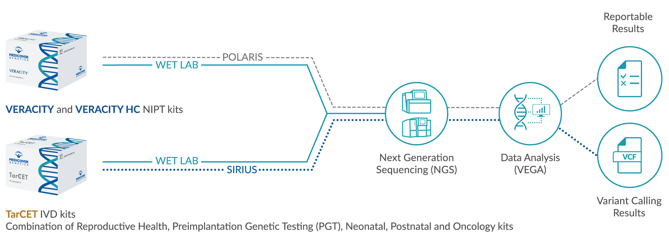
WAYS TO PARTNER WITH US
TarCET IVD Kits
CE-IVD kits containing reagents
for hereditary indications in
an easy-to-use kit form
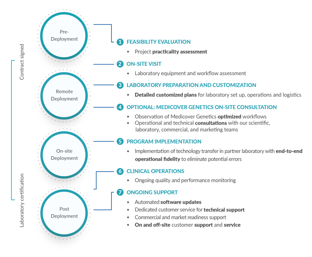
Technology Transfer
Out-of-the-box genetics workflow
from sequencing to reporting
for non-invasive prenatal testing
Contact Us
Please get in touch with us
for any questions, inquiries,
feedback, or with any
comments you might have.
BLOG ARTICLES

We’re thrilled to share the results of the Medicover Genetics essay competition for high school students as well as the two winning essays. This co…
The Human Genome Project, created to determine the sequence of the human genome, was one of the most important biomedical research projects of the 20…
The endometrial microbiome is increasingly recognized as a factor in reproductive health, and imbalances in microbial composition have been linked to…

Spinal muscular atrophy is a rare genetic condition that weakens muscles by affecting the motor nerve cells in the spinal cord. It is a leading genet…

Summer encourages people to spend more time outdoors, soaking in the sun and enjoying the season’s warm weather. While sunlight can have beneficial…

We’re thrilled to share the results of Medicover Genetics essay competition for high school students as well as the two winning essays. This competit…

Cystic fibrosis (CF) is a life-threatening, progressive, inherited condition that causes severe damage to the body, primarily affecting the organs of…

Down syndrome is a genetic condition caused by the presence of an extra copy of chromosome 21. It affects physical growth, facial features, and cogni…

Every year on April 25th, DNA Day celebrates the discovery of DNA’s double helix and the advances we’ve made in understanding genetics. D…

Neurodevelopmental disorders (NDDs) have diverse genetic origins, making diagnosis challenging. A new study analyzing over 1,100 pediatric patients f…